Full Body Thermal Imaging


Thermography, or Digital Infrared Thermal Imaging (DITI) is a totally non-invasive clinical imaging procedure for detecting and monitoring several diseases and physical injuries, by showing the thermal abnormalities present in the body.
Full Body Imaging includes 22-28 views from head to toe.
Upper Body Imaging includes 18-22 views from pubic bone to head.
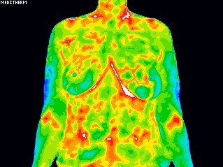
(Female patients: breast study is included with Upper Body or Full Body Thermography at no additional charge).
FEES UP TO
R1700 for Half body
R1900 for Full body
Thermal abnormalities are present in many diseases and physical injuries. Digital Infrared Thermal Imaging (DITI) can help doctors diagnose, evaluate, monitor, and document a large number of injuries and conditions, including soft tissue injuries, sensory/autonomic nerve dysfunction, chronic inflammation, and more. It is used as an aid for diagnosis and prognosis, as well as therapy follow up and rehabilitation monitoring, within clinical fields that include rheumatology, neurology, physiotherapy, sports medicine, oncology, orthopaedics and many others.
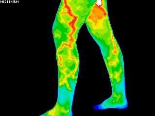
Thermography’s major clinical value is in its high sensitivity to pathology in the vascular, muscular, neural and skeletal systems. It can show a combined effect of the autonomic nervous system and the vascular system, down to capillary dysfunctions. The effects of these changes show as asymmetries in temperature distribution on the surface of the body. Vascular conditions are readily demonstrated including raynauds, vasculitis, limb ischemia, DVT, etc.
Clinical applications for Medical Digital Infrared
Thermal Imaging include
- To define the extent of a lesion of which a diagnosis has previously been made
- To localise an abnormal area not previously identified, so further diagnostic tests can be performed
- To detect early lesions before they are clinically evident
- To monitor the healing process before the patient is returned to work or training

Medical DITI is filling the gap in
clinical diagnosis
- X-ray, CT, Ultrasound and MRI are tests of anatomy
- EMG is a test of motor physiology
- DITI is unique in its capability to show physiological change and metabolic processes. It has also proven to be a particularly useful complementary procedure to other diagnostic modalities.
- Medical DITI is non-invasive. There is no contact, no radiation and no use of contrast agents.
- Medical DITI can offer considerable financial savings by avoiding the need for more expensive investigations.
- Results obtained with medical DITI systems are totally objective and show excellent correlation with other diagnostic tests.
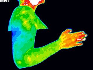
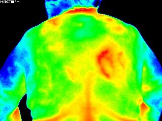
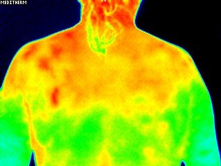
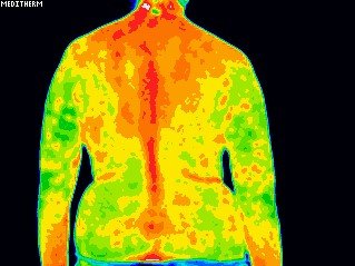
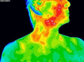
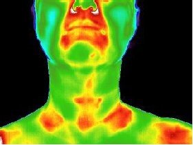
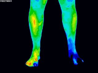
Medical DITI is a noninvasive diagnostic technique that allows the examiner to visualise and quantify changes in skin surface temperature. An infrared scanning device is used to convert infrared radiation emitted from the skin surface into electrical impulses that are visualised in colour on a monitor. This visual image graphically maps the body temperature and is referred to as a thermogram. The spectrum of colours indicates an increase or decrease in the amount of infrared radiation being emitted from the body surface. Since there is a high degree of thermal symmetry in the normal body, subtle abnormal temperature asymmetry’s can be easily identified.
The neuro-thermography application of DITI measures the somatic component of the sympathetic nervous system by assessing dermal blood flow. The sympathetic nervous system is stimulated at the same anatomical location as its sensory counterpart and produces a somatic sympathetic response’. The somatic sympathetic response appears on DITI as a localised area of altered temperature with specific features for each anatomical lesion.
The mean temperature differential in peripheral nerve injury is 1.5°C. In sympathetic dysfunction’s (RSD / SMP / CRPS) temperature differentials ranging from 1° C to 10° C depending on severity are not uncommon. Rheumatological processes generally appear as ‘hot areas’ with increased temperature patterns. The pathology is generally an inflammatory process, i.e. synovitis of joints and tendon sheaths, epicondylitis, capsular and muscle injuries, etc.
Both hot and cold responses may co-exist if the pain associated with an inflammatory focus excites an increase in sympathetic activity. Also, vascular conditions are readily demonstrated by DITI including Raynauds, Vasculitis, Limb Ischemia, DVT, etc.
- Breast Screening for Risk factors and pain
- Unexplained pain
- Headache
- Autoimmune conditions
- Dental and periodontal disease
- Vascular disease (including occlusive disease, deep venous thrombosis)
- Back pain and back injuries
- Identification of myofascial trigger points and tender points
- Infectious and Inflammatory conditions
- Neurological disorders
- Stroke screening and many more